“Our finding that a new cell type is involved in cardiac conduction may lead to better understanding of normal heart function. What really surprised me was that macrophages can depolarize—change their electric charge—when coupled to a heart muscle cell. Down the line, this work on the role of macrophages in conduction may lead to new treatments for cardiac arrhythmias,” said corresponding author Matthias Nahrendorf, HMS associate professor of radiology at Mass General.
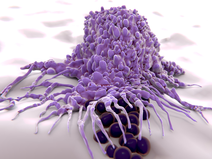
Best known for their role in the immune system, macrophages are primarily responsible for engulfing and digesting microbes, damaged cells and foreign substances. They are found in tissues throughout the body and have recently been shown to have additional functions related to the tissues where they reside. While macrophages are required for healing damaged tissues in the heart, their presence within healthy heart muscle suggests a role in normal heart function. Nahrendorf’s study was designed to investigate their potential role in transmitting and coordinating the electrical signals that stimulate heart muscle contraction.
Initial experiments in mice revealed that cardiac macrophages are more abundant in the atrioventicular (AV) node—a key structure connecting the atria (upper chambers) to the ventricles (lower chambers)—which coordinates contraction timing for the upper and lower chambers. Similarly high concentrations of macrophages were found in AV nodes from human autopsy samples. Subsequent animal experiments found that macrophages connect to heart muscle cells via gap junctions—pore-like structures known to coordinate heart muscle contractions—and that the shifts in electric charge that carry the conduction signal are synchronized between macrophages and adjacent heart muscle cells called myocytes.
Mice lacking a key, gap-junction protein showed an abnormal slowing of signal conduction through the AV node, and a complete depletion of tissue macrophages led to the development of AV block—a delay in conduction between the atria and ventricles that, in human patients, requires pacemaker implantation. Overall, the findings suggest that cardiac macrophages are essential participants in the cardiac conduction system and that changes in their numbers or properties may contribute to heart rhythm abnormalities.
Nahrendorf and his colleagues are continuing to explore the role of macrophages in both the healthy heart and in common disorders of signal conduction. He adds that the cells’ natural propensity to surround and take up materials for disposal could be used to induce macrophages to ingest drugs carried on nanoparticles.
They study was supported by the National Institutes of Health (grants NS084863, HL128264, HL114477, HL117829, HL092577, HL105780 and HL096576). Mass General has filed a patent application covering the work described in this paper.